Quick diagnosis leads to quick treatment.
At Suffolk Animal Hospital, we are able to process our digital X-rays quickly, allowing us to work towards a diagnosis for your pet when speed really counts.
With the advances in digital X-ray technology, we can now manipulate the digital images we take of a pet’s systems to see what is wrong. This has allowed us to detect things like hairline fractures and orthopedic conditions that were previously not visible. We share these digital images with specialists who consult with us on difficult cases.
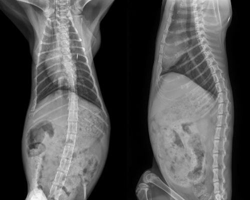
Radiology (X-rays) is routinely used to provide valuable information about a pet’s bones, gastrointestinal tract (stomach, intestines, colon), respiratory tract (lungs), heart, and genitourinary system (bladder, prostate). It can be used alone or in conjunction with other diagnostic tools to provide a list of possible causes for a pet’s condition, identify the exact cause of a problem or rule out possible problems.
How does it work?
When a pet is being radiographed, an X-ray beam passes through its body and hits a piece of radiographic film. Images on the film appear as various shades of gray and reflect the anatomy of the animal. Bones, which absorb more X-rays, appear as light gray structures. Soft tissues, such as the lungs, absorb fewer X-rays and appear as dark gray structures. Interpretation of radiographs requires considerable skill on the part of the veterinarian.

